USU Utilizes State-of-the-Art MRI Technology to Advance Traumatic Brain Injury Research
By Vivian Mason
Mild traumatic brain injury (mTBI)―more commonly known as concussion―has been called the “signature injury” of the wars in Iraq and Afghanistan, affecting a significant number of military personnel due to mission-related injuries from blast exposures or motor vehicle accidents. Though these mTBIs may not be life-threatening, they can produce a wide variety of symptoms that may impact how a person thinks, feels, acts, learns, and sleeps. In a subset of patients, these symptoms can even become chronic conditions.
diagnose subtle changes associated with mTBI, and as a result, medical research currently has an incomplete understanding of what causes these symptoms that affect millions of people in the United States.
That’s where the Uniformed Services University (USU) comes in.
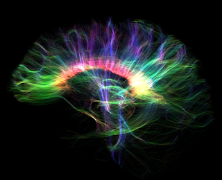
Given the scope of the problem, a high-level USU research team has received funding from a Department of Defense (DoD) grant to investigate new imaging biomarkers for characterization of the underlying pathophysiology in mTBIs in collaboration with GE Research, and GE’s novel, ultra-high-performance Microstructure Anatomy Gradient for Neuroimaging with Ultrafast Scanning (MAGNUS) MRI system. MAGNUS will allow researchers to study the brains of military and civilian personnel who have recently sustained mTBIs.
Dr. Vincent Ho, professor and chair of the Department of Radiology and Radiological Sciences at USU is leading USU’s study efforts. Ho is also the director of research for the Department of Radiology at Walter Reed National Military Medical Center (WRNMMC). The GE Research portion of the study is headed by Dr. Thomas Foo, chief scientist in the Biology and Applied Physics Group at GE.
In March 2020, USU/WRNMMC became the first site with a MAGNUS research system after installing an ultra-high-performance gradient coil insert into a GE 3.0 Tesla clinical MRI scanner. Current clinical MRI systems cannot readily image the brain’s microstructures with sufficient detail to diagnose subtle changes associated with mTBI.
MAGNUS offers much higher gradient strength and slew rate (the change of voltage or other electrical quantity per unit of time) up to 200 millitesla per meter (mT/m) and 500 Tesla per meter per second (T/m/s) versus conventional MRI scanners, which generally offer up to 80 mT/m and 200 T/m/s, a notable difference. This is helpful for detailed microstructural imaging in the setting of mTBI and possibly in Alzheimer’s disease, mild cognitive impairment, or other neuropsychiatric disorders.
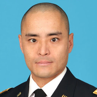
Army Col. (Dr.) Robert Shih―assistant professor and vice chair of the Department of Radiology and Radiological Sciences at USU, as well as chief of Neuroradiology and MRI at WRNMMC―is working with a team of USU researchers to utilize the MAGNUS MRI system for imaging data acquisition and analysis. Shih and his team believe MAGNUS will make a big difference in mTBI research.
Shih says that working on this study provides his team with a unique, exciting opportunity to utilize cutting-edge technology, but that’s certainly not the only reason they’re driven toward this research.
“Our research team at USU is motivated by two things,” Shih explains. “The first is our patients. We want to help our patients and our clinicians better understand what is going on in the concussed brain, which may lead to better management strategies.”
Ongoing research into the mechanisms of mTBI may clear up much of the mystery that still surrounds the cause of symptoms affecting millions of Americans, leading to more effective diagnosis, management, and treatment.
“The second,” Shih concludes, “is a love of technology and all the incredible advances we have seen in radiology over the last few decades, many of which we now take for granted. Our sponsorship by the Department of Defense and our collaboration with GE Research is allowing us to push the envelope of performance at USU.”


