Uniformed Services University Helps Foster Local High School Student Interest in Microscopy and Science
Students from Walt Whitman High School’s microscopy club learned about cutting-edge imaging technology at the Uniformed Services University during a recent visit to the campus.
November 30, 2023 by Hadiyah Brendel
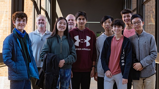
Seven students from Walt Whitman High School, located in Bethesda, Maryland, visited the Uniformed Services University (USU) to get an up-close look at the university’s cutting-edge imaging technology, on November 1st, 2023.
The students are part of the high school’s inaugural microscopy club. Dr. Juanita Anders, professor of Anatomy, Physiology and Genetics (APG) as well as Neuroscience, in the School of Medicine at USU, sponsored the student’s visit to USU’s Biomedical Instrumentation Center (BIC).
Part of the Office of the Vice President for Research, the BIC is a state-of-the-art core facility that houses five suites. The suites – flow cytometry, microscopy, structural biology, histopathology, and genomics – support teaching and research at USU.
Army Col (Dr.) Chad Black, director of the BIC, says the center combines “subject matter experts and research technology together so students and investigators from all aspects of USU” can access a wide sweep of capabilities needed to complete their research protocols and publications in military health care.
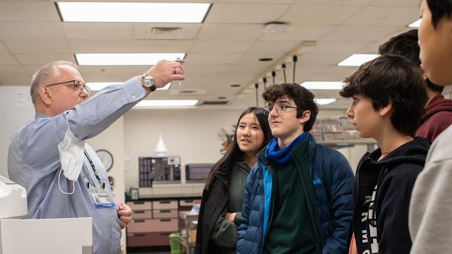
Two of those subject matter experts, Drs. Dennis McDaniel and Fritz Lischka, led the tour.
 |
Dr. Fritz Lischka, a contract employee of the Henry M. Jackson Foundation, is a senior microscopist at BIC and helped lead the tour. (Photo credit: Alex Anders) |
McDaniel is director of Microscopic Imaging at the BIC and a research assistant professor in USU’s Department of Microbiology and Immunology. Lischka, a contract employee of the Henry M. Jackson Foundation for the Advancement of Military Medicine, is senior microscopist at BIC and an adjunct research assistant professor in APG.
Tours of the facility are usually reserved for new faculty candidates or Department of Defense dignitaries visiting the University. The Microscopy Club’s visit was the first of its kind.
“It was kind of surprising,” Anders says of when the club’s president first contacted her.
Black says the club “wanted to see some of the best microscopy in the world. USU has it. It's in the form of the Biomedical Instrumentation Center’s Microscopy Core.” According to Black, ZEISS, the primary manufacturer of BIC’s imaging instruments, says the BIC has probably one of the most cutting-edge microscope suites on the eastern seaboard.
McDaniel and Lischka collaborate with the research community at USU, assisting in a wide range of research domains. First, they discuss what goals a research project aims to achieve with the imaging systems. Then, McDaniel and Lischka provide tiered support. At a minimum, they discuss which microscope is best suited for the project and train the researcher on how to use the microscope. Other times, McDaniel or Lischka will provide more active collaboration and perform image acquisition and image analysis themselves.
“We offer any level of support that the Principal Investigator feels comfortable with,” says Lischka.
McDaniel says “we see things like cancer, parasites, viruses, traumatic brain injury, radiation injury recovery. We see it all, really.”
Two of the instruments demonstrated to the students were the Light Sheet Z1, a lightsheet microscope used to image large, transparent samples in 3D. They also viewed the JEOL JEM-1011 transmission electron microscope, which gives the highest magnification of any microscope.
Anders says the imaging presentation awed the group. One chaperone commented that it was “the most beautiful chromosome she had ever seen in her life” after looking at one of the images. A student remarked how their biology class tomorrow would be “downhill from here,” reports Anders.
Although just formed this September, the Walt Whitman High School Microscopy Club already has 15 students.
“Our objective is to create a platform in which club members can explore the world of microscopy while sharing knowledge and collaborating together,” says club president Aditya Parashar.
The visit to USU was part of their goal “to provide hands-on experiences with microscopes to inspire a shared sense of wonder as we explore function and morphology in biology,” per the club’s mission statement.
In addition to taking an exclusive look at the imaging suite, the tour group also spent time discussing basic principles of microscopy. And though the club members were a bit shy, and perhaps overwhelmed at first, McDaniel says they soon warmed up and were quite engaged.
“I was really kind of looking forward to having the high schoolers come in,” says Lischka. “They are at the very beginning, two months into their microscopy club. So I thought, ‘this is great. You can really get them excited about doing [imaging]’” he added.
For other students interested in microscopy, McDaniels recommends finding someone who works in the field. “I was interested in microscopes and telescopes when I was kid,” he says. In graduate school, he realized he was more interested in the research lab’s heavy focus on microscopes and instrumentation. And he turned it into a career from there.
Lischka adds that no matter what medical research field you may enter, such as molecular biology, oncology, or cell physiology, you can always ask “what benefit could we have from also adding some microscopic approaches?”
While no set plans exist for the club to return next year, Anders and Litschka hope to see some of the students return in the summer. USU hosts a Summer Research Training Program each year, bringing high school students with an interest in science, engineering, or medical career fields to conduct research at the university.
 |
| Walt Whitman High School's Microscopy Club (Photo credit: Alex Anders) |


